


Research and publish the best content.
Get Started for FREE
Sign up with Facebook Sign up with X
I don't have a Facebook or a X account
Already have an account: Login

Bioscience News - GEG Tech top picks
8.8K views |
+0 today
 Your new post is loading...
 Your new post is loading...
Your new post is loading...
BigField GEG Tech's insight:
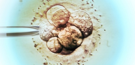
Researchers have captured the most-detailed images yet of human embryos developing in real time, using two common laboratory tools — fluorescent dyes and laser microscopes. Non-invasive imaging approach could lead to innovations in embryo screening.

BigField GEG Tech's insight:
The world's largest stem cell society this week signaled a willingness to reconsider a long-standing restriction on laboratory efforts to grow and study human embryos. In new guidelines, the International Society for Stem Cell Research (ISSCR) also spotlights a possible alternative to using embryos that might be less ethically fraught: emerging methods to model stages of human development with stem cells. ISSCR's influential guidelines previously put the culture of human embryos beyond 14 days postfertilization in its most restrictive category three: "prohibited research activities." The new guidelines, drafted by a task force of scientists and ethicists, omit longer embryo culture from this category and encourage a public discussion about allowing it.
BigField GEG Tech's insight:
The Dutch government is to change the law on embryos to allow them to be grown for ‘very specific’ scientific research under ‘strict conditions’ in an effort to help people unable to have children.
BigField GEG Tech's insight:
Researchers at the University of Cambridge have managed to reconstruct the early stage of mammalian development using embryonic stem cells, showing that a critical mass of cells – not too few, but not too many – is needed for the cells to being self-organising into the correct structure for an embryo to form.
|
As a human embryo grows, a set of molecules directs cells as they multiply and take on specific identities and spatial positions within the embryo.
BigField GEG Tech's insight:
The researchers found ways to recreate a simplified version of gastrulation in a dish by starting with a layer of induced pluripotent stem (iPS) cells, meaning they can differentiate to become any cell type in the body. Next, the scientists added a protein called BMP4, a key signaling molecule in gastrulation, which causes the cells in the box to begin forming the three layers of cells present in the embryo. All cells appear to receive the same BMP4 signal, however, some transform into one cell type while others become different cell types. When creating a gastrulation model, researchers observed that iPS cells contain proteins that are the building blocks of tight junctions. They also noted that tight junctions do not always assemble, and that tight junctions between adjacent cells appear to render cells impervious to BMP4 signals. To confirm the importance of tight junctions in gastrulation, the researchers used CRISPR genome-editing technology to suppress the production of TJP1, a protein crucial for tight junction formation in iPS cells. When they applied BMP4 to cells lacking the TJP1 protein, every cell was activated, not just the peripubic cells. This discovery forms the basis of a new method for efficiently producing these unique cells.
BigField GEG Tech's insight:
Some companies offer tests that rank embryos based on their risk of developing complex diseases such as schizophrenia or heart disease. Are they accurate — or ethical?
BigField GEG Tech's insight:
An initial look at dynamic processes during early human development through 3D cellular imaging.

Single-Cell RNA-Seq Reveals Lineage and X Chromosome Dynamics in Human Preimplantation Embryos
BigField GEG Tech's insight:
Researchers at Karolinska Institutet and the Ludwig Cancer Research in Stockholm, Sweden have conducted a detailed molecular analysis of the embryo's first week of development. Their results show that there are considerable differences in embryonic development between humans and mice, which is the most common organism of study in this field. The authors envision broad utility of this transcriptional atlas in future studies on human development as well as in stem cell research.
BigField GEG Tech's insight:
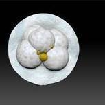
This technology improves the microscopic observation of the embryo. It allows to analyze in 3D the human embryo generated in vitro in full compliance with ethical rules.
|


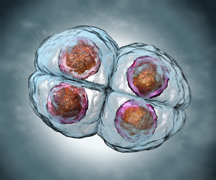


In vitro embryo models supported by methods development in adjacent fields have revolutionized our understanding of embryogenesis.