


Research and publish the best content.
Get Started for FREE
Sign up with Facebook Sign up with X
I don't have a Facebook or a X account
Already have an account: Login

Bioscience News - GEG Tech top picks
8.8K views |
+0 today
 Your new post is loading...
 Your new post is loading...
Your new post is loading...
BigField GEG Tech's insight:
Scientists from the University of Washington recently reported a relatively simple method that would allow ordinary laboratory microscopes to illuminate many of these cellular structures quickly and efficiently. They did not modify microscopes to boost resolution. Instead, they used an approach to swell the tiny, complex structures within cells, bringing them within range of a microscope's existing resolving range. 
BigField GEG Tech's insight:
For the first time, biochemists from the University of Zurich have succeeded in elucidating the structure of the transportation channel inside the nuclear pores in high resolution using high-performance electron microscopes.
|

BigField GEG Tech's insight:
For the first time, the hepatitis C could be observed microscopically by a team of French researchers.
BigField GEG Tech's insight:
In this study, the authors combines intravital microscopy, microscopic X-ray computed tomography and three-dimensional electron microscopy to captured single tumor cells in the vasculature of the cerebral cortex and in subcutaneous tumors, providing unique insights into metastatic events. The versatility of this workflow allows envisioning broad applications in various fields of biological research such as cancer or developmental biology.
|

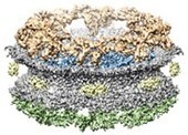


Using 3D electron microscopy, structural biologists from the University of Zurich succeeded in elucidating the architecture of the lamina of the cell nucleus at molecular resolution for the first time.