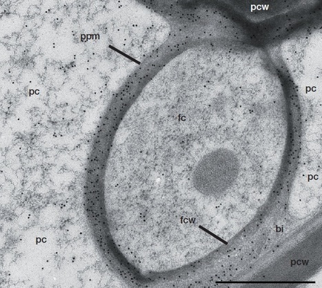
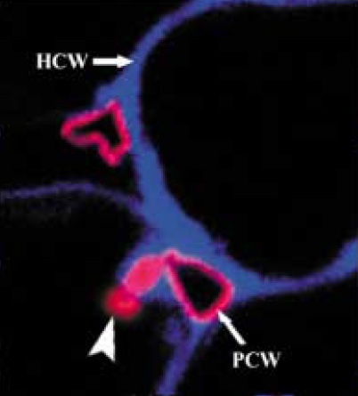
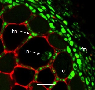
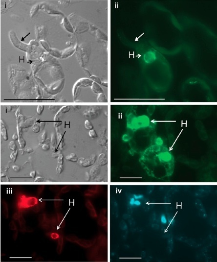
Scooped by
Kamoun Lab @ TSL
onto Host Translocation of Plant Pathogen Effectors |
Get Started for FREE
Sign up with Facebook Sign up with X
I don't have a Facebook or a X account
Your new post is loading...
Kamoun Lab @ TSL's comment,
April 9, 2013 8:08 PM
Two issues came up in our internal discussions of the maggie RFP-NLS assay:
1/ It would be nice to see a decrease of nuclear signal as you move away from the IH/BIC plant cell. Sometimes there are several cells with stained nuclei - one would expect the signal to become weaker and weaker as the protein gets diluted. Some clear documentation of the weakening of the signal as you move away from the first infected cells would be nice. 2/ A question arose last year about whether the artificial NLS is actually carrying the effector inside the plant cells since there are papers about NLS sequences acting as cell-penetrating peptides.
Mark Farman's comment,
April 9, 2013 8:53 PM
There is NO evidence that any Magnaporthe effector translocates across host cell membranes independently of pathogen machinery (nor is there such evidence for oomycetes, for that matter). The Ribot paper is seriously and fundamentally flawed. Actually, I would like to retract my use of the word "translocation" in my earlier comment and replace it with "delivery" because I do not believe that Magnaporthe effectors translocate across membranes at all! Figure that one out...
Mark Farman's comment,
April 9, 2013 8:08 PM
This is a dead cell. The cytoplasm of a living cell should be distributed around the cell periphery, See the image from the Khang et al. paper.
Kamoun Lab @ TSL's comment,
April 9, 2013 8:18 PM
Agreed. Easy to generate such images. See comment in Bozkurt et al. Curr Opin Plant Biol http://kamounlab.dreamhosters.com/pdfs/COPB_2012.pdf
Mark Farman's comment,
April 9, 2013 9:29 PM
There is another serious problem with the experiment shown in this figure: In the legend, the authors state that GUS is inactive in the apoplast and they provide a reference. However, the referenced paper deals with GUS that is expressed in plant cells and directed to the apoplast through the secretory pathway. In this case, the GUS is inactivated by glycosylation in the plant ER. Unfortunately, this cannot possibly explain the lack of GUS staining with the apoplastic SP-7A_GUS fusion protein because the SP-RXLR-EER-GUS protein is perfectly visible and had to navigate the same Phytophthora secretory system. In other words, the negative control did not work as it should have done. End result: experiment is invalid.
|
Gregor Langen's comment,
April 11, 2013 3:57 AM
I don't expect big differences in onion skin response to pathogenic or mutualistic symbionts regarding nuclear movemnt. Eveln mechanical stimulation causes movemenr of the nucleus: Local mechanical stimulation induces components of the pathogen defense response in parsley. Gus-Mayer et al. PNAS 1998 95(14):8398-403.
But I agree that DAPI staining would have helped to clarify identitiy of the structures visible. And yes, halos can be seen
Gregor Langen's comment,
April 11, 2013 4:02 AM
and I agree that a red halo itself doesn't proof that its identical to the nucleus.
Mark Farman's comment,
April 27, 2013 10:50 PM
Even if the onion nuclei are beneath the appressoria, the authors have not established that the fluorescence is in the nucleus, as opposed to being on the surface of the cuticle.
Mark Farman's comment,
April 9, 2013 8:25 PM
Despite the authors assertion that there is no labeling of uninfected cells - there is. Therefore, the fluorescence in the cytoplasmic rings is probably background. Problem is there were no negative (non infected, pre-immune serum) controls in this paper, so we can't tell either way. The fluorescence patterns in the middle panel on the righthand side do not coincide with the cytoplasmic boundaries, indicating a problem with fixation. Most likely protein leached out from the haustoria into the spaces that should be vacuolar.
Mark Farman's comment,
April 9, 2013 8:13 PM
This staining is NOT in the cytoplasm - it is in the extrahaustorial matrix. Also where's the evidence that the stained structure on the bottom right is a nucleus?
Eric Kemen's comment,
May 3, 2013 11:27 AM
I agree, it’s not easy form this picture to see that there is a nucleus underneath the fluorescence signal if one hasn’t seen the DIC picture. But have a look at figure 3 of the same paper where an independent antibody was used in combination with a nuclear stain, to show colocalization of RTP1p signal and DNA staining. In addition, immuno electron microscopy after high pressure freezing shows a signal for RTP1p localization in the cytoplasm (figure 2).
Kamoun Lab @ TSL's comment,
May 17, 2013 10:11 AM
Not much mention of nuclear localization in this follow-up paper http://sco.lt/5jAe01
|