Longevity science
87.1K views | +0 today
Follow
Longevity science
Live longer in good health and you will have a chance to extend your healthy life even further
Your new post is loading...
Scooped by Ray and Terry's
Scoop.it!

Guiding stem cells into damaged hearts with MRI and ultrasonics | KurzweilAI

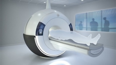
Stem-cell therapy for damaged hearts is a brilliant idea whose time has not yet come. The problem: no way to ensure against faulty initial placement of the stem cells.

Stanford’s Sam Gambhir, PhD, MD, who heads Stanford medical school’s Department of Radiology may have found a way around it.

“You can use ultrasound to visualize the needle through which you deliver stem cells to the heart. But once those cells leave the needle, you’ve lost track of them,” he said.


No comment yet.
Scooped by Ray and Terry's
Scoop.it!

Pill-sized device provides rapid, detailed imaging of esophageal lining

Pill-sized device provides rapid, detailed imaging of esophageal lining | Longevity science | Scoop.it

Physicians may soon have a new way to screen patients for Barrett's esophagus, a precancerous condition usually caused by chronic exposure to stomach acid. Researchers at the Wellman Center for Photomedicine at Massachusetts General Hospital (MGH) have developed an imaging system enclosed in a capsule about the size of a multivitamin pill that creates detailed, microscopic images of the esophageal wall.


The system has several advantages over traditional endoscopy.


No comment yet.
Scooped by Ray and Terry's
Scoop.it!

GE Silent Scan turns down the volume on MRI scanners

GE Silent Scan turns down the volume on MRI scanners | Longevity science | Scoop.it

GE Healthcare has introduced a new data acquisition technology designed to improve patient comfort by largely eliminating the horrible noise generated during an MRI scan. Conventional MRI scanners can generate noise levels in excess of 110 dBA (creating a din that sounds like a cross between a vehicle's reverse warning horn and a Star Trek phaser) but GE says its new Silent Scan MRI technology can reduce this to just above background noise levels in the exam room.

The noise that MRI scanners produce is related to changes in the magnetic field that allow the slice by slice body scan to be carried out. In recent years, industry efforts to speed up the scanning process have also resulted in louder and louder scans. The designers have attempted to dampen these noises with mufflers and baffles, achieving only limited success.


No comment yet.
Scooped by Ray and Terry's
Scoop.it!

Soft squishy robots could replace pill cameras and invasive endoscopes

Soft squishy robots could replace pill cameras and invasive endoscopes | Longevity science | Scoop.it

A magnetic capsule robot may replace endoscopy thanks to work being done at Carnegie Mellon University's Nanorobotics lab.


The lab has received funding to develop a squishy robotic capsule that can be controlled while inside the body. The capsule could replace invasive endoscopes by performing camera imaging, drug injection, tissue sampling, and more.


FDA-approved pill cameras have been in use since 2001, but they can only perform imaging, and move through the body naturally. Robotic devices would have the ability to to stop, back up, deliver drugs, or perform biopsies.

No comment yet.
Scooped by Ray and Terry's
Scoop.it!

Newly developed nanoparticles shine from deep within biological tissue

Newly developed nanoparticles shine from deep within biological tissue | Longevity science | Scoop.it
Newly-developed nanoparticles are able to emit light through up to three centimeters of biological tissue.


Scientists have developed photoluminescent nanoparticles that are able to shine through over three centimeters (1.2 inches) of biological tissue. If attached to anomalies deep beneath the skin, the nanoparticles could allow those anomalies to be seen more clearly from the outside.

No comment yet.
Scooped by Ray and Terry's
Scoop.it!

Firefly gene makes mice glow to track cancer and aging in real time | KurzweilAI

University of North Carolina Lineberger Comprehensive Cancer Center researchers have developed a strain of mice that turns on a gene from fireflies to provide a visual indication of aging and tumor growth in mice. The mice light up whenever another mouse gene, p16INK4a (p16) is is activated (in cells undergoing senescence, the p16 gene is switched on).


No comment yet.
Scooped by Ray and Terry's
Scoop.it!

Compact radiation source could put an X-ray scanner in your pocket

Compact radiation source could put an X-ray scanner in your pocket | Longevity science | Scoop.it

While we’ve seen developments that could see T-ray spectrometers featuring in a future handheld tricorder-like device, good ol’ X-rays could also get a guernsey thanks to an engineering team from the University of Missouri. The team has invented an accelerator about the size of a stick of gum that can create X-rays and other forms of radiation, opening up the possibility of cheap and portable X-ray scanners.


Ray and Terry's 's insight:

Enabling technologies such as inexpensive genomic sequencing, portable medical equipment, and smart-phone medical gadgets can democratize medical care... if the developments are handled properly.

No comment yet.
Scooped by Ray and Terry's
Scoop.it!

Brain scan can sort dementia by type

Brain scan can sort dementia by type | Longevity science | Scoop.it

Scientists say they have found a way to distinguish between different types of dementia without the need for invasive tests, like a lumbar puncture.


US experts could accurately identify Alzheimer's disease and another type of dementia from structural brain patterns on medical scans, Neurology reports.

Currently, doctors can struggle to diagnose dementia, meaning the most appropriate treatment may be delayed.


More invasive tests can help, but are unpleasant for the patient.


No comment yet.
Scooped by Ray and Terry's
Scoop.it!

Hand-held 3D scanner could simplify medical imaging

Hand-held 3D scanner could simplify medical imaging | Longevity science | Scoop.it

Engineers have developed a hand-held scanning device that provides real-time three-dimensional images of the insides of patients’ bodies.


The scanner utilizes optical coherence tomography (OCT), which has been described as “optical ultrasound,” in that it uses reflected light – as opposed to reflected sound – to image internal structures.


No comment yet.
Scooped by Ray and Terry's
Scoop.it!

Web apps for bioinformatics | KurzweilAI

Web apps for bioinformatics | KurzweilAI | Longevity science | Scoop.it
This snapshot is the result of using an Imagejs module to determine how fast brain cancer cells are growing.


A University of Alabama at Birmingham (UAB) team has developed ImageJS, a free app system that analyzes tissue images.


ImageJS is the first in a serie

No comment yet.