Your new post is loading...

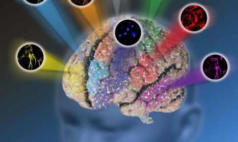

Researchers at Columbia University have made a significant step toward breaking the so-called "color barrier" of light microscopy for biological systems, allowing for much more comprehensive, system-wide labeling and imaging of a greater number of biomolecules in living cells and tissues than is currently attainable. The advancement has the potential for many future applications, including helping to guide the development of therapies to treat and cure disease.
In a study published online April 19 in Nature, the team, led by Associate Professor of Chemistry Wei Min, reports the development of a new optical microscopy platform with drastically enhanced detection sensitivity. Additionally, the study details the creation of new molecules that, when paired with the new instrumentation, allow for the simultaneous labeling and imaging of up to 24 specific biomolecules, nearly five times the number of biomolecules that can be imaged at the same time with existing technologies.
"In the era of systems biology, how to simultaneously image a large number of molecular species inside cells with high sensitivity and specificity remains a grand challenge of optical microscopy," Min said. "What makes our work new and unique is that there are two synergistic pieces - instrumentation and molecules - working together to combat this long-standing obstacle. Our platform has the capacity to transform understanding of complex biological systems: the vast human cell map, metabolic pathways, the functions of various structures within the brain, the internal environment of tumors, and macromolecule assembly, to name just a few."
All existing methods of observing a variety of structures in living cells and tissues have their own strengths, but all are also hindered by fundamental limitations, not the least of which is the existence of a "color barrier."
Via Mariaschnee, Dr. Stefan Gruenwald