 Your new post is loading...
 Your new post is loading...
How can we achieve interlaboratory standardization of flow cytometry assays?
Over the past decade, various liquid biopsy techniques have emerged as viable alternatives to the analysis of traditional tissue biopsy samples. Such surrogate ‘biopsies’ offer numerous advantages, including the relative ease of obtaining serial samples and overcoming the issues of interpreting one or more small tissue samples that might not reflect the entire tumour burden. To date, the majority of research in the area of liquid biopsies has focused on blood-based biomarkers, predominantly using plasma-derived circulating tumour DNA (ctDNA). However, ctDNA can also be obtained from various non-blood sources and these might offer unique advantages over plasma ctDNA. In this Review, we discuss advances in the analysis of ctDNA from non-blood sources, focusing on urine, cerebrospinal fluid, and pleural or peritoneal fluid, but also consider other sources of ctDNA. We discuss how these alternative sources can have a distinct yet complementary role to that of blood ctDNA analysis and consider various technical aspects of non-blood ctDNA assay development. We also reflect on the settings in which non-blood ctDNA can offer distinct advantages over plasma ctDNA and explore some of the challenges associated with translating these alternative assays from academia into clinical use. Advances in circulating tumour DNA (ctDNA) detection and analysis are beginning to be implemented in clinical practice. Nonetheless, much of this development has thus far focused on plasma ctDNA. Theoretically, all bodily fluids, including urine, cerebrospinal fluid, saliva, pleural fluid and others, can also contain measurable ctDNA and can provide several advantages over the reliance on plasma ctDNA. In this Review, Tivey et al. describe the potential roles of ctDNA obtained from non-plasma sources in optimizing the outcomes of patients with cancer.
Application of AFC based assays to confirm infection and predict antimicrobial susceptibility
can deliver actionable results with a performance that meets or exceeds currently
utilised microbiological tests in clinically meaningful timeframes, as demonstrated
for PD peritonitis.
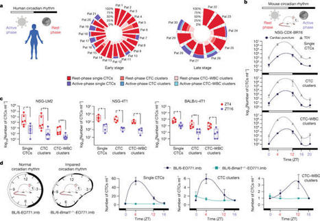
The metastatic spread of cancer is achieved by the haematogenous dissemination of circulating tumour cells (CTCs). Generally, however, the temporal dynamics that dictate the generation of metastasis-competent CTCs are largely uncharacterized, and it is often assumed that CTCs are constantly shed from growing tumours or are shed as a consequence of mechanical insults1. Here we observe a striking and unexpected pattern of CTC generation dynamics in both patients with breast cancer and mouse models, highlighting that most spontaneous CTC intravasation events occur during sleep. Further, we demonstrate that rest-phase CTCs are highly prone to metastasize, whereas CTCs generated during the active phase are devoid of metastatic ability. Mechanistically, single-cell RNA sequencing analysis of CTCs reveals a marked upregulation of mitotic genes exclusively during the rest phase in both patients and mouse models, enabling metastasis proficiency. Systemically, we find that key circadian rhythm hormones such as melatonin, testosterone and glucocorticoids dictate CTC generation dynamics, and as a consequence, that insulin directly promotes tumour cell proliferation in vivo, yet in a time-dependent manner. Thus, the spontaneous generation of CTCs with a high proclivity to metastasize does not occur continuously, but it is concentrated within the rest phase of the affected individual, providing a new rationale for time-controlled interrogation and treatment of metastasis-prone cancers. A study of patients with breast cancer and mouse models demonstrates that most circulating tumour cells are generated during the rest phase of the circadian rhythm, and that these cells are highly prone to metastasize.
Flow cytometry-based Receptor Occupancy can be used to determine saturation at specific time points after dosing to better understand PK/PD relationships, receptor immunomodulation and therapeutic activity.
Webinars On Demand Advances in LED Illumination for Fluorescence Imaging Presenter Kavita Aswani, Senior Biomedical Applications Scientist, Excelitas Technologies Video Duration 1 hour LED illumination for fluorescence microscopy systems has progressed significantly from a time when LED sources had low optical power and a limited range of wavelengths. While the benefits of LED technology drew interest, the limitations prevented large-scale adoption. Advances in the semiconductor industry have now been able to eliminate many of the fundamental hurdles that were previously faced. Today, LED technology is rapidly replacing the lamps that were used in microscopy and fluorescence imaging, including in applications on the extreme ends of the light microscopy spectrum. Xenon lamps have traditionally been relied on by applications that require a light source with uniform spectral stability across the near-UV to visible spectrum. These lamps allow the user to switch filters while maintaining consistent excitation power between wavelengths, which is crucial for quantitative studies. Kavita Aswani, Ph.D. describes the latest advances in illumination for fluorescence imaging, from near-UV to NIR fluorophores. New LED illumination systems are successfully replacing traditional arc lamps in calcium imaging applications. These systems are producing equivalent results with the convenience of LEDs. IR versions of the light sources allow imaging of the popular ICG (indocyanine green) and IR800 dyes. They also provide high signal-to-noise ratios due to the low background in the NIR region. The NIR wavelengths allow for greater depth penetration in thick tissues and living animals. An example of these new systems is the X-Cite NOVEM™ system, which was awarded the prestigious 2021 Microscopy Today Innovation Award. Register below to view this webinar recording to learn more. Photonics Media is the owner and producer of this recording. About the Presenter: Kavita Aswani, Ph.D. is Senior BioMedical Applications Scientist at Excelitas Technologies. She has a doctorate from the University of Iowa and has seven years of scientific research experience and over 20 years of applications experience in the microscopy and fluorescence industry. Aswani has authored and reviewed several publications, as well as journal articles. She has worked in the areas of wide-field illumination microscopy, laser scanning microscopy, and flow cytometry. She is an active member of the Society for Neuroscience and the Royal Microscopical Society. Recording: Advances in LED Illumination for Fluorescence Imaging First Name Last Name Company / Organization Email Address Country - Select Country -USACANABWAFGAGOAIAALAALBANDANTAREARGARMASMATAATFATGAUSAUTAZEBDIBELBENBFABGDBGRBHRBHSBIHBLRBLZBMUBOLBRABRBBRNBTNBVTBWACAFCCKCHECHLCHNCMRCOKCOLCOMCPVCRICUBCXRCYMCYPCZEDEUDJIDMADNKDOMDZAECUEGYESHESPESTETHFINFJIFLKFRAFROFSMGABGBRGEOGHAGINGLPGMBGNBGNQGRCGRDGRLGTMGUFGUMGUYHKGHMDHNDHRVHTIHUNIDNINDIOTIRLISLISRITAJAMJORJPNKAZKENKGZKHMKIRKNAKORKWTLAOLBNLBYLCALIELKALSOLTULUXLVAMACMARMCOMDAMDGMDVMEXMHLMKDMLIMLTMNGMNPMOZMRTMSRMTQMUSMWIMYSMYTNAMNCLNERNFKNGANICNIRNIUNLDNORNPLNRUNZLOMNPAKPANPCNPERPHLPLWPNGPOLPRIPRKPRTPRYPSEPYFQATREUROURUSRWASAUSCGSENSGPSGSSHNSJMSLBSLVSMRSPMSTPSURSVKSVNSWESWZSYCTCATCDTGOTHATJKTKLTKMTLSTONTTOTURTUVTWNTZAUGAUKRUMIURYUZBVATVCTVENVGBVIRVNMVUTWLFWSMYEMZAFZMB Consent Agreement I agree No, I do not agree Consent Agreement Yes, I agree to the storage and use of my information as stated below. No, I do not agree to the storage and use of my information as stated below. PLEASE READ AND INDICATE ABOVE IF YOU AGREE OR IF YOU DO NOT AGREE: Thank you for your interest in Excelitas. Excelitas respects your privacy. Periodically we would like to keep you informed of products, services and developments across the wide array of photonic technologies which you can engage in your applications and systems. To do so, we request your consent to store and utilize your contact information for occasional e-mailings. Please know that Excelitas will never share or sell your contact data. I have read the Declaration of Consent below concerning the storage and use of my data by Excelitas for phone contact, e-mail contact and/or postal contact. Declaration of Consent under Data Protection Legislation I consent to the collection, processing and use of my data by Excelitas Technologies Corp. and its affiliated companies under the condition that such use is limited to providing me with information regarding their products, services, and solutions relevant to my photonics needs or for their customer relations and market research activities.
Congrès annuel conjoints de la Société Française d’Immunologie et l’Association Française de Cytométrie - 22-24 novembre 2022 - Palais des congrès de Nice
Using key flow cytometry parameters, the authors developed a machine learning model to classify acute leukemias and distinguish them from non-neoplastic cytopenias.American Journal of Clinical Pathology...
In the last few decades, the epithelial cell adhesion molecule (EpCAM) has received increased attention as the main membrane marker used in many enrichment technologies to isolate circulating tumor cells (CTCs). Although there has been a great deal of progress in the implementation of EpCAM-based CTC detection technologies in medical settings, several issues continue to limit their clinical utility. The biology of EpCAM and its role are not completely understood but evidence suggests that the expression of this epithelial cell-surface protein is crucial for metastasis-competent CTCs and may not be lost completely during the epithelial-to-mesenchymal transition. In this review, we summarize the most significant advantages and disadvantages of using EpCAM as a marker for CTC enrichment and its potential biological role in the metastatic cascade.
Metastasis, tumor relapse, and development of resistance to treatment are responsible for most cancer-related mortality. Various laboratories, including... 21 comments on LinkedIn
Results of a novel pilot study using the CellSearch Circulating Endothelial Cell assay to analyze vascular dysfunction in patients with systemic sclerosis
Quels laboratoires continueront de réaliser des LDT après la fin du marathon #IVDR 2022 ?
Les laboratoires cliniques qui utilisent des test
Circulating tumor cells (CTCs) are cells that are shed from tumors into the bloodstream. Cell enrichment and isolation technology as well as molecular profiling via next-generation sequencing have allowed for a greater understanding of tumor cancer biology via the interrogation of CTCs.
|
Scientific Article | Here we report a protocol for the quantification and differentiation of myocardial B-lymphocytes based on their location in the...
One of the challenges medicine faces is the constantly growing resistance of pathogens to various classes of antibiotics.In this study, we investigated the use of capillary electrophoresis (CE) to characterize and assess the physiological states of three clinical bacterial strains - Staphylococcus...
Since its development in the 1960s, flow cytometry (FCM) was quickly revealed a powerful tool to analyse cell populations in medical studies, yet, for many years, was almost exclusively used to analyse eukaryotic cells.
Cyanobacteria are Gram-negative oxygen-producing photosynthetic bacteria that are useful in the pharmaceutical and biofuel industries. Monitoring of oxidative stress under fluctuating environmental conditions is important for determining the fitness, survival, and growth of cyanobacteria in the...
Malaria diagnosis based on microscopy is impaired by the gradual disappearance of experienced microscopists in non-endemic areas.Aside from the conventional diagnostic methods, fluorescence flow cytometry technology using Sysmex XN-31, an automated haematology analyser, has been registered to suppo...
Three-dimensional (3D) fluorescence imaging is important to accurately capture and understand biological structures and phenomena. However, because of its slow acquisition speed, it was difficult to implement 3D fluorescence imaging for imaging flow cytometry.
Frontiers in Flow Cytometry™ is an annual event that will bring together cutting-edge flow cytometry and cell-sorting research applications from scientists across the globe.
This 24-hour event will feature keynote presentations, live and on-demand webinars, demos, networking opportunities and more.
Register now to join us!
Discover the events that have occurred over the course of history that mark the evolution of flow cytometry.
Impacts potentiels du règlement UE IVDR sur le laboratoire de cytométrie en flux clinique Dr Maurizio Suppo Comme pour beaucoup d'autres idées visant à améliorer les produits et procédures existants, le chemin du nouveau règlement européen relatif aux dispositifs médicaux de diagnostic in vitro...
We optimized a procedure for quantitative expression profiling of surface antigens on blood leukocyte subsets.The workflow, bioinformatics pipeline and optimized flow panels enable the following: 1) mapping the expression patterns of HLDA-approved mAb clones to CD markers; 2) benchmarking new antib...
Since the SARS-CoV-2 virus emerged as a threat to public health, numerous methods have emerged, or have been re-deployed, for studying the virus, and for introducing novel drugs and vaccines to combat the pandemic.
T and natural killer (NK) cells are effector cells with key roles in anti-HIV immunity, including in lymphoid tissues, the major site of HIV persistence. However, little is known about the features of these effector cells from people living with HIV (PLWH), particularly from those who initiated...
|
 Your new post is loading...
 Your new post is loading...


